The Cardiothoracic Surgery Research Laboratory is focused on developing surgical treatments for cardiothoracic conditions, including atrial fibrillation, and translating them into clinical practice.
Established over 40 years ago, our lab conducts innovative research backed by multiple sources of funding, including an R01 grant from the NIH. The lab has two main areas of research focus:
- Surgical treatment of cardiac arrhythmias and rhythm management
- Ablation technology
The Cardiothoracic Surgery Research Laboratory is led by Christian Zemlin, PhD, MSc, whose work focuses on the mechanisms and treatment of arrhythmias. His research has been funded by the American Heart Association, the NIH, intramural funding and industry.
Zemlin earned his master’s degree in physics from the Technical University of Berlin and his doctorate in theoretical physics from Humboldt University in Berlin in 2002. He completed his postdoctoral research in cardiac electrophysiology at SUNY Upstate Medical University in Syracuse.
His research uses voltage-sensitive fluorescent probes to experimentally study cardiac activity, and computer modeling to understand how arrhythmias are initiated and maintained. Zemlin developed a new ablation modality for cardiac tissue based on ultrashort electric pulses that cause irreversible electroporation.
Principal investigator
Christian W. Zemlin, PhD, MSc
Associate Professor of Surgery and Biomedical Engineering
Section of Cardiac Surgery
Division of Cardiothoracic Surgery
Director, Cardiothoracic Surgery Research Laboratory
Contact
Christian Zemlin, PhD, MSc
Clinical Sciences Research Building
Email: [email protected]
Phone: 314-362-8300
Opportunities
Apply for a training position in our lab
Areas of research focus
Surgical treatment of cardiac arrhythmias and rhythm management
For the past four decades, our laboratory has conducted studies to develop surgical treatments for cardiac arrhythmias and translated them into clinical practice.
Our earliest efforts were directed at arrhythmias associated with ischemic heart disease, Wolff-Parkinson-White syndrome, and atrioventricular node reentry, and resulted in clinical interventions routinely used today.
For each arrhythmia, we initially defined the fundamental substrates and mechanisms in animal experiments.
The surgical approach was worked out first in the laboratory and then perfected in clinical studies to account for the differences between animal and human arrhythmia mechanisms.
The validity of this innovative approach has been borne out by the long-term success and adoption of these interventional approaches both by surgeons and electrophysiologists.

Our work has led to the development of the most successful single treatment for atrial fibrillation (AF), the Cox-Maze procedure. In the Cox-Maze procedure, lines of scar are created on the atria to disrupt potential reentrant pathways.
Our laboratory also spearheaded the effort to simplify the Cox-Maze procedure by replacing the surgical incisions with lesions created using thermal ablation technology, which made the procedure easier to perform with less morbidity while preserving its high efficiency.
The Cox-Maze procedure has become the gold standard for the treatment of AF, and it is the only surgical procedure to have received FDA approval.”
In a propensity-matched study of patients with AF undergoing heart surgery, a concomitant Cox-Maze procedure resulted in a 10-year survival rate of 62%, compared to 42% for patients who did not receive a Cox-Maze procedure.
Ablation technology
In the original Cox-Maze procedure, lesions were created by cutting and sewing tissue. To simplify the procedure, cryoablation and radiofrequency ablation were introduced. Both methods are thermal: Cryoablation cools the tissue to kill it while radiofrequency ablation heats the tissue until it dies.
Our laboratory has tested every major cryoablation and radiofrequency ablation device that has been used clinically, as well as alternative technologies such as focused ultrasound and microwave ablation that were never widely adopted clinically.
Currently, a new ablation approach called pulsed field ablation (PFA) is in clinical testing. PFA uses strong electric fields to electroporate the membranes of cells. The figure on the right illustrates PFA ablation of an atrial appendage. The ablation clamp closely resembles those used in radiofrequency ablation.

Current research
Chronic severe mitral regurgitation and atrial fibrillation
Mitral regurgitation (MR) is the most common valvular disease in humans. Many MR patients later develop atrial fibrillation. To understand the underlying mechanisms, we developed an animal model of MR, based on avulsing some of the chordae that hold the mitral valve in place.
After chordae have been avulsed, MR can be demonstrated in a transthoracic echocardiogram (see Panel A), and the valve shows coaptation defect (Panel B). After MR creation, left atrial volume increased dramatically, by about 200% over 9.5 months.
We are now using this model of MR to understand the molecular mechanisms of atrial and ventricular remodeling with the goal of developing interventions to prevent atrial fibrillation and other secondary diseases.
We are also studying the effects of mitral regurgitation in patients referred for surgery with without atrial fibrillation. We are using novel, noninvasive imaging technology, in attempt to define the underlying substrate in atrial fibrillation. We hope to use this information in the future to guide surgical ablation.


Bipolar radiofrequency ablation
An important treatment option for patients with atrial fibrillation is the ablation of cardiac tissue to create nonconducting lesions that disrupt the pathways of reentrant arrhythmias. In current clinical practice, cardiac ablation is performed thermally, either by heating tissue with radiofrequency currents or by freezing it with a cryogen. We are regularly testing new thermal ablation devices for their ability to create transmural lesions.

Nanosecond pulsed field ablation of cardiac tissue
Nanosecond pulsed field ablation (PFA) is an alternative to the thermal ablation methods described above. PFA is nonthermal and relies on strong electric fields to disrupt cell membranes in order to ablate tissue. PFA can significantly reduce ablation times while providing excellent ablation depth. It also avoids thermal effects such as char buildup on the ablation clamp that can lead to incomplete lesions for radiofrequency ablation.

Optogenetics and sonogenetics in the heart
The beating of the cardiomyocytes that make up the heart is synchronized by electrical signals that are generated by the sinus node and propagate over the heart. If the heart is genetically modified to include light-sensitive ion channels, activation can be achieved with a light (optogenetics). Similarly, adding ultrasound-sensitive ion channels allows stimulation with ultrasound (sonogenetics).
In collaboration with Drs. Chao Zhou, Jianmin Cui, and Hong Chen from the Department of Biomedical Engineering, we are developing murine models of optogenetics and sonogenetics in the heart, enhancing the options for non-invasive stimulation of the heart.

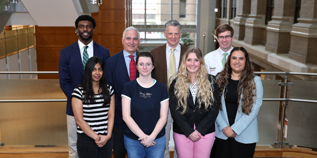
Our team
Ralph J. Damiano Jr., MD
Evarts A. Graham Professor of Surgery
Section of Cardiac Surgery
Division of Cardiothoracic Surgery
- Phone: 314-362-7260
Matthew R. Schill, MD
Assistant Professor of Surgery
Section of Cardiac Surgery
Division of Cardiothoracic Surgery
- Phone: 314-362-7260
Christian W. Zemlin, PhD, MSc
Associate Professor of Surgery and Biomedical Engineering
Section of Cardiac Surgery
Division of Cardiothoracic Surgery
Director, Cardiothoracic Surgery Research Laboratory
Fellows
- Jack Yi, MD
- Ruth Obiarinze, MD
Staff
- Lab supervisor: Samantha Procasky, MS
- Veterinary technician III: Kristen Barth
Emeritus investigator
News and updates
Abstract accepted for STS annual meeting 2025

Dr. Jack Yi’s abstract “Impact of Tricuspid Regurgitation on Outcomes after Cox-Maze IV Procedure” was accepted for a poster presentation at the STS Annual Meeting from January 24-26th in Los Angeles, California.
Annual pool party and Dr. Yu send off

Dr. Jakraphan Yu completed his one-year research fellowship with our lab in June 2024. We sent him off with our annual pool party and he received a framed certificate from Drs. Damiano and Zemlin. We wish him the best for his work at Vajira Hospital in Bangkok, Thailand.
Drs. Schill and Zemlin receive pilot grant from the Department of Surgery

Drs. Schill and Zemlin received a pilot research award from the Department of Surgery. They are principal investigators on the proposal “Endovascular Catheter-Based Application of Electroporation”, which was submitted jointly with Dr. Zayed from Vascular Surgery and Genin from Mechanical Engineering. The award is $50k over one year, with the goal of developing the project to the point at which it can secure federal funding.
Yu receives 2024 Kouchoukos Award

Dr. Jakraphan Yu (center) from our lab receives the Kouchoukos award from Drs. Kouchoukos (left) and Kreisel (right)
Lab alumni
- Meghan Kelly, MD
- Ali Khiabani, MD
- Joshua Manghelli, MD
- Martha McGilvray, MD
- Robert McGregor, MD
- Tariana Yates, MD
- Jakrakphan Yu, MD