Breast cancer is a significant health concern worldwide, and early detection is crucial for improving survival rates. Mammograms have long been the gold standard for breast cancer screening, but other methods are also available.
Understanding mammograms
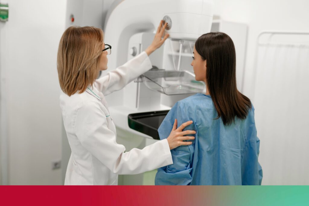
Mammograms are specialized X-ray images of the breast used to detect abnormalities. There are two main types of mammograms: screening and diagnostic.
- Screening mammograms: These are routine tests performed on women without any symptoms to detect breast cancer early. They typically involve taking two X-ray images of each breast.
- Diagnostic mammograms: These are more detailed X-rays used when there is a suspicion of breast cancer, such as a lump or unusual symptoms. They may involve additional images and angles.
During a mammogram, the breast is compressed between two plates to spread the tissue and obtain clear images. While this compression can be uncomfortable, it is necessary for accurate results.
Learn more about breast cancer>>
Benefits of mammograms
- High sensitivity: Mammograms are highly sensitive and can detect tumors that are too small to be felt.
- Early detection: They are effective in identifying early-stage breast cancer, which is crucial for successful treatment.
- Widely accessible: Mammograms are available in most health care facilities and are recommended by major health organizations.
Did you know?
Researchers at WashU Medicine developed a new technology using AI to analyze mammograms and improve the accuracy of predicting a woman’s risk of breast cancer.
Other breast cancer screening methods
While mammograms are the most common screening tool, other methods can also be used, especially for women with dense breast tissue or those at high risk.
Ultrasound
Ultrasound uses sound waves to create images of the breast tissue. It is often used as a supplementary tool to mammograms.
- How it works: A handheld device called a transducer is moved over the breast, emitting sound waves that bounce off tissues and create images.
- When it is used: Ultrasound is typically used to further evaluate abnormalities found in mammograms or for women with dense breast tissue.
- Benefits and limitations: Ultrasound can distinguish between solid masses and fluid-filled cysts but may not detect all cancers.
MRI (Magnetic Resonance Imaging)
MRI uses magnetic fields and radio waves to produce detailed images of the breast.
- How it works: The patient lies face down on a table with their breasts positioned in openings. The table slides into the MRI machine, which takes images.
- When it is used: MRI is often used for high-risk women, such as those with a strong family history of breast cancer or genetic mutations.
- Benefits and limitations: MRI is highly sensitive and can detect cancers that mammograms might miss, but it is more expensive and can lead to false positives.
Clinical breast exam (CBE)
A clinical breast exam is a physical examination of the breasts performed by a health care professional.
- How it is performed: The health care provider uses their hands to feel for lumps or abnormalities.
- Benefits and limitations: CBE is a simple and low-cost method but is less effective than imaging techniques in detecting early-stage cancers.
Self-examination
Self-examination involves women checking their own breasts for lumps or changes.
- Importance of self-awareness: Regular self-exams can help women become familiar with their breasts and notice any changes.
- How to perform a self-exam: Women should use their hands to feel for lumps and visually inspect their breasts for changes.
- Benefits and limitations: While self-exams can help detect changes, they are not a substitute for professional screening methods.
Comparing mammograms with other screening methods
Each screening method has its strengths and weaknesses, and the choice of method depends on individual risk factors and preferences.
Effectiveness in detecting breast cancer
- Mammograms: Highly effective for early detection, especially in women over 50.
- Ultrasound: Useful for dense breast tissue but may miss some cancers.
- MRI: Highly sensitive but prone to false positives.
- CBE and self-exams: Less effective for early detection but important for overall breast health awareness.
Accuracy and reliability
- Mammograms: High accuracy, especially with digital technology.
- Ultrasound and MRI: High accuracy but can lead to overdiagnosis.
- CBE and self-exams: Variable accuracy depending on the examiner’s skill.
Accessibility and cost
- Mammograms: Widely available and often covered by insurance.
- Ultrasound and MRI: More expensive and less accessible.
- CBE and self-exams: Low cost (or free) and widely accessible.
Suitability for different risk groups
- Mammograms: Recommended for women over 40 or those at average risk.
- Ultrasound and MRI: Recommended for high-risk women or those with dense breast tissue.
- CBE and self-exams: Suitable for all women as part of regular health check-ups.
Breast cancer screening is vital for early detection and successful treatment. While mammograms remain the gold standard, other methods like ultrasound, MRI, and clinical exams play essential roles in comprehensive breast health.
Women should consult their health care providers to determine the best screening strategy based on their individual risk factors and preferences. Regular screenings and awareness are key to combating breast cancer and ensuring a healthier future.
To schedule a screening, please visit the Siteman Cancer Center website or call for an appointment with a surgical oncologist at WashU Medicine: 314-362-2280.